Arman Moshaveri1*, Daryoush Babazadeh2, Faezeh Modarresi-Ghazani3, Veghar Hejazi4, Muhammad Saeed5, and Pouria Ahmadi Simab6
- Faculty of Veterinary Medicine, Karaj Branch, Islamic Azad University, Karaj, Iran
- School of Veterinary Medicine, Shiraz University, Shiraz, Iran
- Drug Applied Research Center, Tabriz University of Medical Sciences, Tabriz, Iran
- MD, Tabriz University of Medical Sciences, Tabriz, Iran
- College of Animal Science and Technology, NW A&F University, Yangling, Shaanxi, China
- Faculty of Veterinary Medicine, Sanandaj Branch, Islamic Azad University, Sanandaj, Iran
* Corresponding author: Arman Moshaveri, Faculty of Veterinary Medicine, Karaj Branch, Islamic Azad University, Karaj, Iran. Email: armanvetmoshaveri@gmail.com
ABSTRACT
Introduction: Nitroglycerin can increase the Cycle Guanosine Mono Phosphate level, enhance nitric oxide rate in tissues dilate vessels, and intensify perfusion within tissues. The aim of the present study was to conduct a microscopic investigation addressing the effect of topical 2% Nitroglycerin ointment on wound healing in rabbits.
Materials and methods: Six adult male New Zealand white rabbits, weighing approximately 2.25 kg were used. Two wounds were created on each side of the spinal column. The wounds reached the deep fascia and their dimensions were 15 15 mm. The left wound was used as the control and the right wound was used as the experimental one. Immediately after the creation of wounds, a layer of nitroglycerin 2% with 1 mm thickness was put on the experimental wound daily for seven days. On days 3, 5, 7, and 14 after cutting, both the experimental and control wounds with a margin of healthy tissues were taken for the histopathological examination.
Results: The distance of the two edges on the wound in treated wounds reached a significant difference, compared with control wounds on day 14. The number of inflammatory cells (with neutrophils format) in the treatment group was significantly less than those of the control group starting from day 5. In addition, the fibrin clot diameter in the treatment group was significantly less than the control group on days 5, 7, and 14. At the beginning of the experiment angiogenesis in the control group was more than in the treatment group, but it was the same in both groups on day 7, and angiogenesis in the treatment group was more than in the control group after day 7. The volume of granulation in the treatment group was more than control group and there was a significant difference on days 5, 7, and 14. Epithelial tissues diameter was higher in the treatment group and the difference became significant on day 14.
Conclusion: In conclusion, the findings indicated a promising function of topical NTG in wound healing of anal fissures, tendinopathies, CNH, diabetic foot, or skin flap necrosis.
Keywords: Histopathology, Nitroglycerin, Rabbit, Wound Healing
Introduction
Wound healing stimulation is one of the proposed issues in medical and veterinary medicine. Different physical and chemical procedures have been proposed to heal the wound and which has been investigated and regularly updated as an important issue. Perfusion is one of the most important factors in wound healing procedure, so the medicines which increase perfusion to the wound can have a positive effect on this procedure1,2.
Nitroglycerin(NTG) known as 1,2,3-trinitro propane is one of the medicines which enhances Nitric Oxide (NO) rate in tissues, dilates vessels, and intensifies perfusion within the tissues by an increase in the Cycle Guanosine Mono Phosphate (CGMP) level2,3. Topical NTG and lidocaine significantly increase radial artery diameter (RAD) within 30 to 60 minutes with no effect on the contralateral radial artery or blood pressure, indicating a direct local effect on the radial artery4. Moreover, this medicine prohibits calcium channels activities to change the calcium level intracellularly5.
A great deal of basic and clinical research on the physiologic and pathophysiologic roles of NO in cardiovascular function has been conducted since Endothelium-derived Relaxing Factor (EDRF) was found to be NO6. Furthermore, intracellular calcium ion has an important role on the overall process of wound healing. These findings were applied in previous studies to indicate the role of calcium in human wound healing5.
Topical nitroglycerin ointment (NTGO) would be absorbed through the skin and complete its effectiveness which last for 2-12 hours. NTC has a very short half-life (1-4 min in humans) and it would be metabolized in the liver7.
Some limited investigations have been conducted on the topical effects of NTG on the healing of anal fissures and the success rate of skin graft which lead to contradicted results8,9. The effect of honey and NTG on wound healing was investigated10. The findings revealed that the wound healing period in honey and NTG groups was significantly shorter than the control group, and also, NTG could increase the blood flow of the wound site, and consequently accelerated the healing process, however, it took some time for its effects to appear. Heggers et al.11 compared the effect of Aloe Vera and NTG on wound healing and found that Aloe Vera was more effective. The effects of Sildenafil and NTG on skin flaps of rats after nicotine application were investigated, the results indicated that cotreatment with oral sildenafil and local NTG can improve skin survival12. In another study, Dunn and Brodland13 examined the effect of this medicine on 88 patients with surgical wounds but no significant effects on skin graft and flap enhancement were reported. On the other hand, during an investigation on rats’ intestinal anastomotic healing14, it was mentioned that NTC was an effective medicine. Furthermore, in a study by Hwang et al.15 the positive effects of NTG on hemorrhoidectomy and wound healing were confirmed. In an investigation Sanei et al.9, they compared glyceryl trinitrate (GTN) with diltiazem for the treatment of chronic anal fissure and it was concluded that both were equally effective and might be preferred to become the first line in treating chronic anal fissures. Kim et al.16 evaluated the efficacy of combining NTGO and Adipose-Derived Stem Cells (ADSCs) as a superior wound healing treatment and reported that wound-healing rate was accelerated in the NTGO-applied wound. Karanik et al.17 also assessed the effects of GTN on promoting wound healing after hemorrhoidectomy and the findings were indicative of the GTN effectiveness in the treatment within three postoperative weeks.
The meta-analysis of NTGO use after hemorrhoidectomy as an analgesic and wound healing accelerator has been focused in a study18. It was reported that GTN had a significant pain-relieving effect lasting three to seven days postoperatively and significantly improved wound healing at three weeks. The aim of the current study was to investigate the histopathological effects of NTG 2% on wound healing of rabbit skin.
Materials and Methods
Animals
Six male adult New Zealand white rabbits aged 8-9 months, weighing approximately 2 kg were included in this study. The rabbits were kept in individual cages with equal environmental conditions and diet. They had 12 hours of light and 12 hours of darkness, also the temperature and humidity were kept at 22 2°C and 50 5% respectively. The rabbits were fed standard food pellets (Razi institute, Iran) starting two weeks before the investigation.
Wound cutting method
Two wounds were created on each side of the spinal column of all animals. The wounds reached to the deep fascia and their dimensions were 15 15 mm. The left wounds were used as control and the right wounds as the experimental wounds.
Nitroglycerin application
Immediately after cutting the wounds a layer of NTG 2% (Berenguer-Infale, Germany) in vaseline (Tehran Chemi Company, Iran) base with 1 mm thickness was used on the experimental wounds daily for seven days, the control wounds received a layer of vaseline with the same thickness.
Histopathological investigation
On days 3, 5, 7, 14, 21, 28 after cutting, 10 samples with a margin of healthy tissues from both experimental and control wounds of a rabbit on each day of sampling were taken for histopathological examination. The samples were fixed in 10% neutral formalin solution and dehydrated with a series of ethanol solutions. The samples were also processed by the conventional paraffin embedding method. Microtome sections were prepared at 5 thicknesses, stained with Hematoxylin and Eosin (H & E), and observed under a light microscope (Olympus, Japan). Histopathological assessments were made based on the observations, the distance between the wound edges measurement, infiltration rate of inflammatory cells, the diameter of a fibrin clot, the number of blood vessels, the volume of granulation, and the diameter of newly formed epithelial tissue.
Statistical analysis
The data recorded from both experimental and control wounds were evaluated and analyzed by comparing the means in SPSS software (version 22). Duncan test was used to investigate the significant differences at ¬¬level of p ≤ 0.05.
Results and Discussion
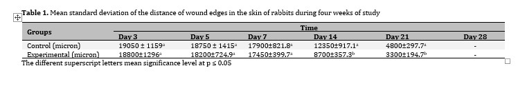
In the current study, the effects of topical NTG on the process of wound healing were assessed using histopathological investigations. The results indicated that the distance of wound edges, an important indicator of wound healing, became significantly shorter 14 days after starting the treatment (p ≤ 0. 05, Table 1). This is important because the shorter the wound edges are, the sooner the wound will shrink and start healing.
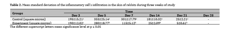
In addition, the infiltration of inflammatory cells significantly decreased from the fifth day (p ≤ 0.05, Table 2).
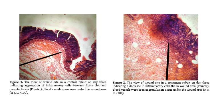
Comparing the microscopic images of control and treated wounds on the third day, it was obvious that the inflammatory cells decreased in the treated wound while there was an aggregation of these cells in the control wound (Figures 1 and 2).
A lower infiltration of inflammatory cells is an indicator of faster inflammation clearance and consequently, faster healing. The histologic findings also reported that angiogenesis was at a similar level at the beginning of the treatment, while on the fifth day, there was severe angiogenesis in the control group and newly formed vessels in the treatment group (Figures 3 and 4).
This trend was reversed when the number of blood vessels formed in the treatment group started increasing until it reached a significant level on day 14 (Table 4). In fact, the medicine causes the blood vessels to become more stable until the complete treatment of the wound in the treatment group is achieved. The Higher vessel formation and its continuity until the end of the treatment improved the transfusion of healing factors and accelerated healing. As the treatment with topical NTG continued, a better organization of collagen and limitation of the wound was observed in the treatment group, compared to the control group (Figures 5 and 6). Fibrin clot average diameter was lower in the treatment group at the beginning of treatment and it reached a significant level on day 5 (p ≤ 0.05) and remained that way by the end of the experiment (Table 3). This means that there is a negative relationship between granulation and fibrin clot diameter, so the less the fibrin clot diameter, the more the granulation volume resulting in faster healing. Epithelial tissue formation had started on day 14 in the treatment group and day 21 in the control group, and it was higher in the treatment groups compared to the control group on days 14, 21, and 28 (p ≤ 0.05, Table 6, Figures 7 and 8). The more the epithelial tissues cause faster wound healing with respect to the studies conducted on chronic fissure healing on 50 people. Newly formed granulation was additionally higher in the treatment group from the beginning of the experiment and significant from day 5 (p ≤ 0.05, Table 5). The comparison between granulation of samples is demonstrated in Figures 9–12. Finally, the epithelial tissue diameter was higher in the experiment group, compared to the control group on day 14, and remained that way until day 28. The difference was significant on days 14 and 21 (Table 6). According to the present literature, the effect of topical NTG has been efficient in wound healing8,9,19. In a recent study, Gündüz et al.20 investigated the effects of NTG in the stasis zones of a burning model and reported significant differences in edema, inflammation, and vascular proliferation in the NTG 2% group. Additionally, it was indicated NTG 2% could increase vascular proliferation in the zones of stasis and improve wound healing. In another study on rats, the effect of topical NTG together with Aloe vera was investigated on a rat model of diabetic foot. The results indicated that this combination could yield faster healing of the ulcer21. Moreover, another meta-analysis by Liu et al.22 on 12 trials with 1095 patients of hemorrhoidectomy reported that using topical NTG had beneficial effects on pain relief and wound healing. This result together with the results of the meta-analysis by Ratnasingham et al.18 provides strong evidence of NTG as a wound-healing agent. The topical NTG 2% was effective in the treatment of both the symptoms and lesional appearance of chondrodermatitis nodularis helicis (CNH) in a non-invasive manner23.