Introduction
Unlike coccidiosis which is a disease caused by parasites (Apicomplexa phylum and the Eimeriidae family), infectious bursal disease (IBD) is a viral infection that affects young domestic chickens all over the world. Depression, watery diarrhea, ruffled feathers, and dehydration are all symptoms of the clinical condition. In young chicks, the disease can manifest as a clinical or subclinical disease, depending on the infectious bursal disease virus (IBDV) strain and the existence of maternal immunity. All pathogenic IBDVs induce lesions in the bursa of fabricious in both clinical and asymptomatic phases of the illness. The cloacal bursa might expand and develop a yellowish transudate on its surface. Hemorrhages of the serosal and mucosal services have been reported on occasion. Around 7-10 days after infection, the bursa atrophy, which involves the loss of B-lymphocytes, begins. Immunosuppression is linked to the loss of B-lymphocytes, but immunosuppression and secondary infections are more common in birds who recover from the condition. Immunosuppression severity is determined by the virulence of the infecting virus and the host’s age.
Etiology and Transmission
The infectious bursal disease is caused by a birnavirus, which is most easily isolated from Fabricius’ bursa but may also be isolated from other organs. It is excreted in the feces and spread by fomites from home to house. It is a tough bug to get rid of because it’s so steady.
IBDV has been divided into two serotypes. In hens, serotype 1 viruses cause illness, and antigenic diversity between strains is possible. This antigenic diversity is mostly due to antigenic drift, although antigenic changes can also be caused through genome homologous recombination. The virus’s serotype 2 strains infect hens and turkeys but do not induce illness or immunosuppression in these animals. IBDVs have been found in a variety of bird species, including penguins, and IBDV antibodies have been found in a variety of wild avian species. The role of IBDV in disease transmission in these wild birds is unclear.
Clinical Findings
Infectious bursal disease is extremely infectious, and the severity of infection is determined by the chicken’s age and breed, as well as the virus’s virulence. Infections can be either subclinical or clinical in nature. Infections that occur before the age of three weeks are generally asymptomatic. When immature B cells occupy the bursa and maternal protection has gone, chicks are most susceptible to clinical illness around 3–6 weeks of age, but severe infections have occurred in Leghorn hens up to 18 weeks of age.
Because of the financial losses, early subclinical infections are the most important manifestation of the illness. Due to the death of immature lymphocytes in the bursa of Fabricius, thymus, and spleen, they produce severe, long-lasting immunosuppression. The humoral (B cell) immune response is harmed the greatest, whereas the cell-mediated (T cell) immunological response is harmed less. Chickens that have been immunosuppressed by early IBDV infections do not react well to immunization and are more susceptible to infections with viruses and bacteria that are ordinarily nonpathogenic. Infections with IBDV are known to aggravate a variety of disorders. Some IBDV strains can induce subclinical infections in older birds (3–6 weeks old), resulting in feed efficiency losses and prolonged time to market. Immunosuppression is generally only temporary in these circumstances, and convalescent birds can regain most or all of their humoral immune function. Secondary infections that arise as a result of the transitory immunosuppression, on the other hand, might result in severe financial losses.
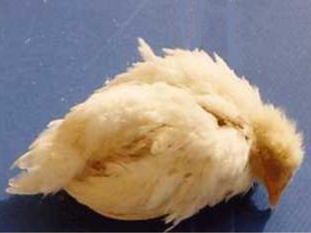
In clinical infections, illness onset occurs after a 3–4 day incubation period. Severe prostration, incoordination, watery diarrhea, dirty vent feathers, vent picking, and cloacal inflammation are all possible symptoms in chickens. Flock morbidity is normally 100%, while death can range from 5% to 60%, depending on the virus strain and chicken breed. Layer chickens have a greater mortality rate than broiler chickens. Recovery takes around a week, and broiler weight growth takes 3–5 days longer. The existence of maternal antibodies alters the disease’s clinical progression.
The virus’s virulence varies greatly amongst field strains. Viruses ranging in severity from naturally attenuated to highly virulent (vv) have been identified. The vvIBDV strains that can cause substantial mortality (>20 percent) were discovered in Europe for the first time. They were discovered in South and Central America in 1999, and in the United States in 2009. They spread over the Middle East, Asia, and Africa, and were first identified in South and Central America in 1999.
Lesions
The lesions revealed during necropsy will vary depending on the strain of IBDV. The cloacal bursa is enlarged, edematous, yellowish, and occasionally hemorrhagic in strains that produce clinical illness, notably in birds who died of the disease. vvIBDV strains induce comparable cloacal bursa lesions, as well as congestion and bleeding in the pectoral and leg muscles. Some IBDV strains can cause cloacal bursa atrophy without causing any visible lesions in the organ. Due to the death and lack of regeneration of the bursal follicles, chickens who have recovered from IBDV infections have tiny, atrophied cloacal bursas.
Diagnosis
- Clinical examination of the cloacal bursa for macroscopic and microscopic lesions, followed by molecular identification of the viral VP2 gene using RT-PCR, can be used to make a diagnosis.
- The IBDV genotype is determined using VP2 gene sequence analysis.
- It is feasible, although not always essential, to isolate viruses in chicken embryos or chicken embryo fibroblast cell cultures.
Gross lesions in the cloacal bursa are used to make the first diagnosis of infected bursal illness. The bursa is next examined under a microscope for lymphocyte depletion in the follicles. The most common method for detecting IBDV in diagnostic samples is to employ molecular diagnostic tests. In bursa tissue, the reverse-transcriptase-PCR technique is utilized to identify the viral genome. The viruses were further classified into genogroups using sequence alignments and phylogenetic analyses of the VP2 coding region. After maternal antibodies have faded, samples for molecular diagnostic testing are routinely taken.
With inocula from birds in the early stages of illness, IBDV can be isolated in 8- to 11-day-old, antibody-free chicken embryos. Inoculation is more sensitive to the chorioallantoic membrane than to the allantoic sac. Cell cultures of chicken embryo fibroblasts, cells from the cloacal bursa, and existing avian and mammalian cell lines may also be used to isolate IBDV strains. IBDV strains that have been modified for cell culture generate a cytopathic effect and can be utilized for viral titration and neutralization experiments.
In convalescent chicks, serology can be utilized to determine the existence of IBDV antibodies. The most common method for quantifying IBDV antibodies is to utilize commercially available ELISA kits. Because most young chicks have maternal antibodies, the presence of IBDV antibodies in chicks is not usually an indicator of infection.
Control
There is no therapy available. After depopulation, thorough decontamination of polluted farmland has had mixed results. At 1–21 days of age, live vaccinations of chicken embryo or cell-culture origin with varied low pathogenicity can be given via eye drop, drinking water, or SC routes. Maternal antibodies can affect vaccination replication and consequently the immunological response, while more virulent vaccine strains can override greater levels of maternal antibody. In ovo or at hatch, vectored vaccinations expressing the IBDV VP2 protein in the herpesvirus of turkeys (HVT) can be employed. Maternal antibodies have no effect on these HVT-IBD vaccinations. Immune-complex vaccinations (live-attenuated viruses coupled to antibodies) are also available for in ovo or hatch delivery.
High maternal antibody levels during early brooding of chicks in broiler flocks (and certain commercial layer farms) can help to prevent early infection, immunosuppression, or both. Breeder flocks should be vaccinated one or more times during the growth season, initially with a live vaccination and then with an oil-adjuvanted, inactivated vaccine soon before egg production. There are inactivated vaccines made from chicken embryos, bursas, or cell cultures. Compared to live vaccinations, these vaccines produce greater, more consistent, and longer-lasting antibody levels. A quantitative serologic test, such as viral neutralization or ELISA, should be used to assess the immunological state of breeder flocks on a regular basis. If antibody levels drop, chickens should be revaccinated to ensure that their offspring have appropriate protection.
The objective of any IBD immunization program should be to employ vaccines that match the antigenic profile of the field viruses as precisely as possible. Diagnostic testing for field strain genetic sequences can be used to determine the best immunization strategy.
Key Points
- Infectious bursal disease is a disease that affects young chickens and is caused by the infectious bursal disease virus. The virus attacks immature B-lymphocytes, causing immunological suppression in convalescent birds, which leads to subsequent infections.
- The virus may be found all over the world, and it’s diagnosed by examining the cloacal bursa clinically and identifying the viral genome molecularly.
- In order to control IBD, breeder flocks are vaccinated to establish maternal immunity in young chicks. Antigenic drift necessitates the use of vaccinations with antigenic structures that closely match the infecting virus. As maternal antibodies diminish, vaccination in ovo or in early chicks with vectored or live-attenuated vaccinations can help enhance protection.