Introduction
Coccidiosis is caused by protozoa belonging to the Apicomplexa phylum and the Eimeriidae family. The majority of Eimeria species infect different parts of the gut in chickens. The infectious phase is quick (4–7 days) and is marked by parasite proliferation in host cells as well as significant intestinal mucosa destruction. Coccidia in poultry are usually host-specific, with various species parasitizing different regions of the gut. Coccidia, on the other hand, may parasitize the whole digestive system in birds such as quail.
Etiology of Coccidiosis
Coccidia is virtually commonly prevalent in poultry-raising operations, but clinical illness develops only after sensitive birds consume significant amounts of sporulated oocysts. Oocysts are shed in the droppings of both clinically sick and recovered birds, contaminating feed, dust, water, litter, and soil. Mechanical carriers may be used to transfer oocysts (eg, equipment, clothing, insects, farmworkers, and other animals). Fresh oocysts are not infectious until they sporulate, which takes 1–2 days under ideal circumstances (21°–32°C with appropriate moisture and oxygen). The prepatent period lasts four to seven days. Depending on the environment, sporulated oocysts might live for a long time.
Host genetics, dietary variables, concomitant illnesses, host age, and coccidium species all influence pathogenicity. Because schizogony occurs in the lamina propria and crypts of Lieberkühn of the small intestine and ceca, respectively, and produces widespread bleeding, Eimeria necatrix and Eimeria tenella are the most pathogenic in chickens. In chukars, E kofoidi and E legionensis are the most pathogenic, whereas, in bobwhite quail, E lettyae is the most pathogenic. Pheasants are susceptible to Eimeria species, notably E phasiani and E colchici. The majority of species grow in epithelial cells that line the villi. In most cases, protective immunity develops in response to moderate and persistent infection. Although true age-immunity does not exist, older birds are typically more resistant to illness than younger birds due to earlier virus exposure.
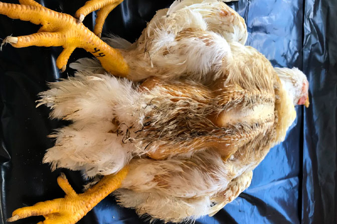
Clinical Findings of Coccidiosis
Coccidiosis symptoms range from slowed development to a large number of clearly unwell birds, severe diarrhea, and significant mortality. The amount of food and water consumed is low. Weight loss, culling, decreased egg production, and higher mortality may occur as a result of epidemics. Subacute infections of intestinal species, which would normally be classified as subclinical, might produce depigmentation and, in the case of Clostridium spp infection, secondary infection. Survivors of severe infections recover in 10–14 days, although lost performance may never be recovered.
The lesions are nearly completely found in the intestine, and they frequently have a unique location and appearance that aids in identification.
Diagnosis
The species present is determined by the location in the host, the development of lesions, and the size of the oocysts. The presence of oocysts in feces or intestinal scrapings can be used to confirm coccidial infections; however, the amount of oocysts present has minimal bearing on the severity of clinical illness. The severity of the lesions, as well as information about the flock’s appearance, morbidity, daily mortality, feed intake, growth rate, and lay rate, are all crucial factors in determining the diagnosis. It is recommended that multiple fresh specimens be necropsied. The classic E tenella and E necatrix lesions are pathognomonic, however infections caused by other species are more difficult to identify. The coccidial species may be distinguished quite well by comparing lesions and other indications to diagnostic charts. Infections with many coccidial species are frequent.
If oocysts, merozoites, or schizonts are found microscopically and lesions are severe, clinical coccidiosis is suspected. It is possible that subclinical infections of coccidiosis are inconsequential, and that poor performance is due to other flock issues.
Control
Coccidial infection cannot be prevented with current therapeutic approaches. Poultry that are kept on wire flooring at all times to keep them away from droppings have fewer illnesses; clinical coccidiosis is infrequent in such situations. Vaccination and anticoccidial medication prevention are two further strategies of control.
Vaccination
After a natural infection, a species-specific immunity develops, the degree of which is mostly dependent on the depth of infection and the frequency of reinfections. T-cells are the main players in protective immunity.
Live, sporulated oocysts of several coccidial species are injected at low dosages in commercial vaccinations. Day-old chicks should be vaccinated with modern anticoccidial vaccinations, either at the hatchery or on the farm. Chickens are reinfected on the farm by descendants of the vaccination strain since the vaccine merely acts to introduce infection. The majority of commercial vaccinations contain live coccidia oocysts that have not been attenuated. Rather than biological attenuation, the self-limiting feature of coccidiosis is exploited as a sort of attenuation for certain vaccinations. Attenuated coccidia lines are included in certain vaccinations offered in Europe and South America. Vaccination of game birds has shown potential in research.
Layers and breeders who are kept on the floor litter need to be protected. Historically, these birds were given an inadequate dose of an anticoccidial medicine during their early development, with the hope that immunity would develop over time as a result of frequent exposure to wild coccidia. Because it’s impossible to regulate all of the elements that impact coccidia reproduction under real-world settings, this strategy has never been totally successful. Vaccination programs are gaining popularity, despite the fact that anticoccidial medications have been chosen for the protection of these birds. The bonuses are different every day. The practicality of vaccination in broilers is improving because of better delivery procedures and a wider selection of coccidia strains in the product.
Anticoccidial Drugs
Anticoccidials are used in the feed to avoid sickness and the financial losses that commonly accompany subacute infection. Because most of the harm occurs before symptoms appear and because medications cannot entirely stop an epidemic, prophylactic usage is preferable. Because of the practical constraints of feed delivery, therapeutic treatments are frequently administered via water. Antibiotics and higher amounts of vitamins A and K are occasionally included in the feed to speed up recovery and avoid recurring illnesses.
Anticoccidial medications can be coccidiostatic, halting intracellular coccidia growth but allowing development to continue after drug removal, or coccidiocidal, killing coccidia throughout their development. When administered short-term, several anticoccidial medications may be coccidiostatic, but when given long-term, they may be coccidiocidal. Coccidiocidal anticoccidials are now employed in chicken production.
During the use of anticoccidials in the feed, the natural development of immunity against coccidiosis may occur. This may be of minimal relevance in the production of broilers during a short growout of 37–44 days. Replacement layer pullets need natural immunity since they are likely to be exposed to coccidial infections for long periods of time after anticoccidial medicines are stopped. Anticoccidial strategies for layer and breeder flocks are designed to protect against acute outbreaks while immunizing infection.
To satisfy regulatory criteria and save production costs, anticoccidials are typically removed from broilers 3–7 days before slaughter. Longer withdrawal increases the danger of coccidiosis outbreaks since broilers’ sensitivity to infection varies at this period.
For confinement-reared turkeys up to the age of 8–10 weeks, a preventative anticoccidial is administered. Outbreaks are thought to be less likely in older birds.
Amprolium is a thiamine antagonist (vitamin B1). Coccidia that divide quickly have a high thiamine demand. When administered at the highest permissible dose in feed (125–250 ppm), amprolium has an 8:1 safety margin. Because amprolium has low efficacy against some Eimeria spp., it has been used with the folic acid antagonists ethopabate and sulfaquinoxaline to broaden its scope. Today, amprolium is mostly used to purify water during clinical epidemics.
Clopidol and quinolines (e.g., decoquinate, methylbenzoquate) impede mitochondrial energy generation, making them coccidiostatic against Eimeria spp. during early growth. Clopidol and quinolines have a wide range of species and are occasionally combined for synergy. During prolonged usage, however, resistance may develop quickly.
Nicarbazin, the first medication with true broad-spectrum action, has been in widespread use since 1955. Although the mechanism of action is unknown, it is assumed to work by inhibiting succinate-linked nicotinamide adenine dinucleotide dinucleotide reduction and the energy-dependent transhydrogenase, as well as calcium buildup in the presence of ATP. Nicarbazin is poisonous to layers and causes mottling of egg yolks, reduced egg output, and brown egg shell blanching. Broilers require a 4-day withdrawal period. In warmer weather, medicated birds are more susceptible to heat stress.
Nitrobenzamides (e.g., dinitolmide) are the most effective coccidiostats against asexual stages. Unless paired with additional products, efficacy is restricted to E tenella and E necatrix.
Robenidine, a guanidine chemical, permits coccidia to grow intracellularly but hinders full schizont production. When used for a short period of time, it is coccidiostatic, and when used for a longer period of time, it is coccidiocidal. During the course of treatment, drug resistance may emerge. To eradicate unfavorable flavor induced by residues in poultry meat, a 5-day withdrawal period is required.
Toltrazuril and diclazuril are effective against a wide range of coccidia. At 1 ppm in the feed, diclazuril is mostly utilized for prevention, whereas toltrazuril is largely used for water purification.
Coccidiosis in different types of Poultry
Turkeys
Only E adenoides, E dispersa, E gallopavonis, and E meleagrimitis are considered pathogenic among the seven species of coccidia found in turkeys. Nonpathogenic species include E innocua, E meleagridis, and E subrotunda. After being expelled from the host, oocysts sporulate within 1–2 days; the prepatent phase is 4–6 days.
The lower ileum, ceca, and rectum are infected with E adenoeides and E gallopavonis. These animals are notorious for causing death. The villi and crypts’ epithelial cells contain the developmental phases. The intestinal wall may be thickened and dilated in the afflicted area. Huge quantities of oocysts can be found in thick, creamy substances or caseous casts in the intestines or excreta. The upper and mid-small intestine are the most commonly infected areas by E meleagrimitis.
Parasites may infect the lamina propria or deeper tissues, resulting in necrotic enteritis (see Necrotic Enteritis). E dispersa causes a creamy, mucoid enteritis that affects the whole intestine, including the ceca, when it infects the upper small intestine. A large number of gametocytes and oocysts are seen in the lesions. Reduced feed consumption, fast weight loss, droopiness, ruffled feathers, and severe diarrhea are all common symptoms in infected flocks. Wet, mucusy droppings are prevalent. Clinical infections are uncommon in poults older than 8 weeks. The rate of morbidity and death might be rather high.
Ducks
both wild and farmed ducks have been shown to carry the most specific coccidia. It has been proven that Eimeria, Wenyonella, and Tyzzeria spp. are present. T perniciosa is a recognized pathogen that causes mucohemorrhagic or caseous material to inflate throughout the small intestine. Eimeria spp. is also known to be pathogenic. Domestic duck coccidia is thought to be rather nonpathogenic in some species. Coccidiosis outbreaks in wild ducklings occur seldom but dramatically in ducklings 2–4 weeks old; morbidity and death may be substantial.
Geese
The most well-known coccidial infection in geese is E truncata, which causes enlargement of the kidneys and the appearance of poorly delimited yellowish-white streaks and patches. Masses of oocysts and urates have dilated the tubules. It’s possible that there will be a lot of deaths. At least five more Eimeria spp. have been found in the intestines of geese, although they are minor parasites.
Chickens
E tenella infections affect just the ceca and are marked by a build-up of blood in the ceca as well as bloody droppings. Cecal cores are accumulations of clotted blood, tissue debris, and oocysts detected in birds that have survived the acute stage.
E necatrix causes significant lesions in the small intestine’s anterior and middle sections. On the serosal surface, little white dots, generally interspersed with spherical, bright- or dull-red patches of varied sizes, can be detected. “Salt and pepper” is a common description for this look. If aggregates of big schizonts can be seen microscopically, the white dots are diagnostic for E necatrix. The intestinal wall thickens and the diseased region dilates to 2–2.5 times its usual width in severe instances. Blood, mucus, and fluid can all fill the lumen. Dehydration can occur as a result of fluid loss. The sexual part of the life cycle is completed in the ceca, despite the damage in the small intestine. Only the ceca contain E necatrix oocysts. Oocysts of different species may be discovered in the region of significant lesions as a result of concurrent infections, deceiving the diagnostician.
The most prevalent source of infection is E acervulina. On gross inspection, there are many white, oval, or transverse patches in the upper portion of the small intestine that may be clearly identified. In most flocks, the clinical course is long and culminates in poor growth, an increase in culls, and a modest rise in mortality.
The lower small intestine, rectum, ceca, and cloaca all contain E brunetti. The mucosa is pale and disturbed in severe infections, but there are no identifiable foci, and it may be thickened. Coagulative necrosis and mucosal sloughing occur throughout the small intestine in severe infections.
E maximum produces dilatation and thickening of the small intestinal wall, petechial bleeding, and a crimson, orange, or pink sticky mucous exudate and fluid. The midgut’s exterior is frequently covered in white pinpoint foci, and the region may seem engorged. The oocysts and gametocytes (especially macrogametocytes) found in the lesions are noticeably enormous.
In the lower small intestine, E mitis is known to be pathogenic. The lesions are ambiguous, however, they may mimic mild E brunetti infections. Finding tiny, spherical oocysts associated with the lesion distinguishes E mitis from E brunetti.
E praecox infects the upper small intestine and causes no visible lesions, although it can slow down development. The oocysts of E acervulina are bigger and more abundant in afflicted locations. It’s possible that the contents of your intestines are fluid. E praecox is regarded as having a lower economic value than the other species.
E hagani and E mivati grow in the small intestine’s anterior section. E hagani’s lesions are vague and difficult to classify. E mivati, on the other hand, can cause severe lesions comparable to E acervulina. E mivati may cause reddening of the duodenum due to villi denudation in severe infections. Some believe these species are of doubtful origin, however, research using genetic diagnostics appears to back them up.
Conclusion
Despite the importance of the chicken sector to each country’s economy, the threat of coccidiosis has been a restricting factor. Though the struggle against chicken coccidiosis has been ongoing for decades, the reappearance of the disease, along with novel Eimeria parasite types in various geographical locations, necessitates a renewed focus on management measures. Anticoccidial medications, vaccinations, and tight management practices have shown to be beneficial, but the rise of genetic and antigenic diversity poses a significant threat to the efficacy of current anticoccidial vaccines and treatments. It is critical to comprehend the dynamic in the development of variety among genes that encode potential vaccine antigens at the genome level, as well as the accompanying processes that drive this diversity, in order to build an effective vaccine.