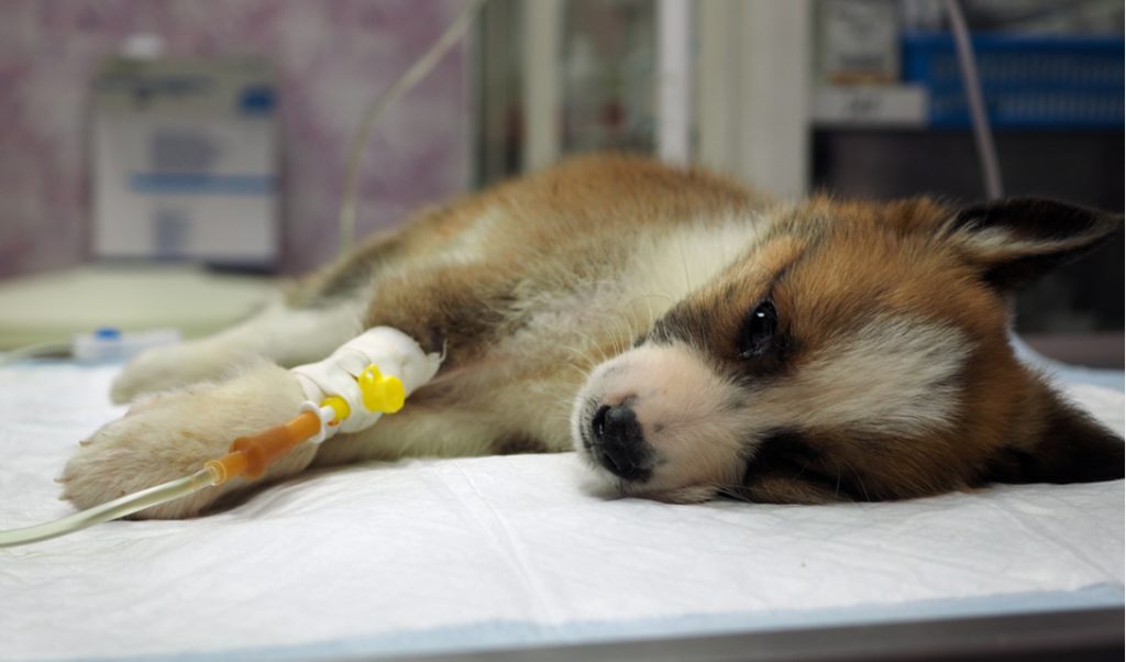
Canine parvovirus (CPV): Clinical Signs, Diagnosis, Treatment and Prognosis, Prevention and Control
Canine parvovirus (CPV) is a highly contagious and the common cause of acute, infectious gastrointestinal sickness in puppies and/or dogs that have not been immunized. Its exact origin is uncertain; however, it is thought to have originated from the feline panleukopenia virus. It’s a single-stranded DNA virus that’s resistant to several common detergents and disinfectants, as well as temperature and pH fluctuations. Infectious CPV can survive inside for at least two months at room temperature; outside, if sheltered from sunshine and desiccation, it can survive for months, if not years. Clinical sickness is generally attributable to CPV-2b in North America; however, infection with a newer and similarly deadly strain, CPV-2c, is becoming more widespread, with at least 15 states reporting cases. To date, no association has been identified between CPV strain and the severity of the clinical disease.
Young (6 weeks to 6-month-old), unvaccinated, or incompletely vaccinated dogs are most susceptible. Breeds described as at increased risk include:
- Rottweilers
- Doberman Pinschers
- American Pit Bull Terriers
- English Springer Spaniels
- German Shepherds
Puppies born to a dam with CPV antibodies are protected from infection for the first few weeks of life assuming adequate colostrum intake; nevertheless, vulnerability to infection rises as maternally acquired antibody wanes. The more severe clinical disease has been linked to stress (e.g., from weaning, overcrowding, starvation, etc.), concurrent intestinal parasitism, or enteric pathogen infection (e.g., Clostridium spp, Campylobacter spp, Salmonella spp, Giardia spp, coronavirus). Intact male dogs are more likely than intact female dogs to develop CPV enteritis in dogs over the age of six months.
Within 4–5 days of exposure (typically before clinical indications appear), the virus is shed in the feces of infected dogs, and for up to 10 days following clinical recovery. Infection is contracted by direct contact with virus-containing feces in the mouth or nose, or indirectly through contact with virus-infected fomites (eg, environment, personnel, equipment). Viral replication begins in the lymphoid tissue of the oropharynx, with systemic disease and hematogenous dispersion following. CPV primarily infects and kills rapidly proliferating cells in the crypt epithelium of the small intestine, lymphopoietic tissue, and bone marrow. Epithelial necrosis, villous atrophy, decreased absorption capacity, and disturbed gut barrier function ensue from the destruction of the intestinal crypt epithelium, with the risk of bacterial translocation and bacteremia.
Lympopenia and neutropenia are caused by the death of hematopoietic progenitor cells in the bone marrow and lymphopoietic tissues and are aggravated by an increased demand for leukocytes throughout the body. Infection in pregnancy, in puppies under the age of eight weeks, or in pups delivered to unvaccinated moms without naturally existing antibodies might cause myocardial infection, necrosis, or myocarditis. With or without indications of enteritis, myocarditis might appear as abrupt cardiopulmonary failure or delayed gradual cardiac failure. Because most bitches have CPV antibodies from immunization or natural exposure, CPV-2 myocarditis is uncommon.
Clinical Signs of Canine parvovirus
Clinical indications of parvoviral enteritis usually appear 5–7 days after infection, however they can appear anywhere between 2 and 14 days. Initial symptoms may be vague (e.g., lethargy, anorexia, fever), but within 24–48 hours, vomiting and hemorrhagic small-bowel diarrhea develop. Nonhemorrhagic diarrhea affects around a quarter of all dogs. Depression, fever, dehydration, and dilated and fluid-filled intestinal loops are all possible physical examination results. Abdominal discomfort should be investigated further to rule out the possibility of intussusception. Animals that have been severely impacted may seem collapsed, with a delayed capillary refill time, poor pulse quality, tachycardia, and hypothermia—signs that might indicate septic shock. Although CPV-related leukoencephalomalacia has been described, CNS symptoms are more usually caused by hypoglycemia, sepsis, or acid-base and electrolyte imbalances. Infection that is undetectable or subclinical is prevalent.
Lesions
- Canine parvovirus can cause the following gross necropsy lesions:
- a darkened and thicker intestinal wall
- intestinal contents that are fluid, mucoid, or hemorrhagic
- edema and congestion of the lymph nodes in the abdomen and thorax
- atrophy of the thymus
- Pale streaks in the myocardium in the case of CPV myocarditis
Intestinal lesions are marked by widespread necrosis of the crypt epithelium, loss of crypt architecture, and villous blunting and sloughing on histological examination. Peyer’s patches, peripheral lymph nodes, mesenteric lymph nodes, thymus, and spleen show lymphoid tissue and cortical lymphocyte depletion, as well as bone marrow hypoplasia. In dogs that died of complications such as acute respiratory distress syndrome, systemic inflammatory response syndrome, endotoxemia, septicemia, pulmonary edema, alveolitis, and bacterial colonization of the lungs and liver may be detected.
Diagnosis of Canine parvovirus
- Signalment, history, and clinical indicators have led to suspicion.
- fecal parvoviral antigen testing or viral PCR confirmation
Any young, unvaccinated, or incompletely vaccinated dog with relevant clinical indications, especially those residing in or freshly obtained from a shelter or breeding kennel, should be suspected of canine parvovirus enteritis. Most dogs acquire a moderate to severe leukopenia, which includes lymphopenia and neutropenia, throughout the course of the disease. A poor prognosis has been linked to leukopenia, lymphopenia, and the absence of a band neutrophil response within 24 hours of initiating therapy.
The serum biochemical profile may reveal prerenal azotemia, hypoalbuminemia (GI protein loss), hyponatremia, hypokalemia, hypochloremia, and hypoglycemia (due to insufficient glycogen stores in young puppies and/or sepsis, potentially a poor prognostic indicator), as well as increased liver enzyme activities. Even with the more recently developed CPV-2c strain, commercial ELISAs for antigen detection in feces are readily accessible and offer good to exceptional sensitivity and specificity.
All animals exhibiting relevant clinical indications should be evaluated right away so that proper isolation protocols may be implemented. The feces of most clinically unwell dogs contain substantial amounts of viruses. Because of the dilutional impact of large-volume diarrhea, false-negative findings can arise early in the course of the disease (before peak viral shedding) or after the fast fall in viral shedding that occurs within 10–12 days of infection (3-4 days after the development of clinical signs). Within 4–10 days of immunization with modified-live CPV vaccine, false-positive findings might be seen.
PCR testing, electron microscopy, and viral isolation are all other options for detecting CPV antigens in feces. A 4-fold rise in serum IgG titer during a 14-day period or the identification of IgM antibodies in the absence of recent (within 4 weeks) vaccination are required for serodiagnosis of CPV infection. This type of testing is hardly employed.
Treatment and Prognosis
· To prevent the spread of canine parvovirus, dogs with the virus should be separated from other dogs as soon as possible.
· Fluid and electrolyte therapy, nutritional support, anti-emetics, and antibiotics are all part of the treatment plan.
Treatment for canine parvovirus enteritis focuses on restoring fluid, electrolyte, and metabolic imbalances as well as preventing subsequent bacterial infection. Oral electrolyte solutions can be given if there is no considerable vomiting. For dogs with moderate to severe dehydration, SC administration of an isotonic balanced electrolyte solution may be sufficient to repair small fluid deficits (5%), but not for dogs with moderate to severe dehydration.
IV fluid treatment with a balanced electrolyte solution is beneficial for the majority of dogs. Dehydration must be corrected, ongoing fluid losses must be replaced, and maintenance fluid requirements must be met in order for therapy to be successful. Hypokalemia and hypoglycemia in dogs must be monitored carefully. If electrolytes and blood glucose levels cannot be checked on a regular basis, IV fluids should be supplemented with potassium (potassium chloride 20–40 mEq/L) and dextrose (2.5–5%).
Colloid treatment should be explored if GI protein loss is significant (albumin 2.0 g/dL, total protein 4.0 g/dL, signs of peripheral edema, ascites, pleural effusion, etc.). Nonprotein colloids (e.g., pentastarch, hetastarch) can be given as boluses (5 mL/kg, maximum 20 mL/kg) during a 15-minute period. The remaining 20 mL/kg maximum dose can be given as a constant-rate infusion over the course of 24 hours, with the volume of crystalloids delivered reduced by 40%–60%. Based on human research, there may be a risk of coagulopathy or acute renal damage while using hydroxyethyl starch solutions. There are few veterinary research available. Transfusion of fresh frozen plasma, on the other hand, may partially restore serum albumin while also giving serum protease inhibitors to reduce the inflammatory response in the body.There is no evidence to support the use of convalescent or hyperimmune serum from dogs recovering from CPV enteritis as a passive immunization method.
Because of the potential of bacterial translocation across the damaged intestinal epithelium and the possibility of concomitant neutropenia, antibiotics are recommended. A beta-lactam antibiotic (e.g., ampicillin or cefazolin [22 mg/kg, IV, three times daily]) will give enough coverage for both gram-positive and anaerobic bacteria. Additional gram-negative coverage (eg, enrofloxacin [5-10 mg/kg/day, IM or IV] or gentamicin [9-12 mg/kg/day, IV]) is needed for severe clinical symptoms and/or significant neutropenia. Antibiotics with aminoglycosides should not be used until dehydration has been treated and fluid treatment has been established. Enrofloxacin has been linked to articular cartilage injury in dogs who are 2–8 months old and should be stopped if joint discomfort or swelling occurs. Cephalosporins of the second or third generation (e.g., cefoxitin, ceftazidime, cefovecin, and others) are particularly worth considering because of their broad range of action against both gram-positive and gram-negative bacteria. Antibiotic treatment is usually only required for a short period of time (e.g., 5–7 days).
Antiemetic treatment is used if vomiting is prolonged, causes dehydration and electrolyte imbalances, or prevents medicines and nutritional assistance from being administered orally. Maropitant (1 mg/kg/day, IV) and ondansetron (0.5 mg/kg, IV, three times daily) appear to be similarly efficient at controlling vomiting in dogs with CPV enteritis. Metoclopramide (0.3 mg/kg, PO or SC, twice day, or 1–2 mg/kg/day as a constant-rate infusion) can be used as both an antiemetic and a prokinetic in dogs with substantial stomach stasis. Despite antiemetic treatment, vomiting may continue. Other causes of vomiting, such as intussusception, should be considered in these circumstances. Anti-diarrheas are not advised since keeping intestinal contents in a weakened gut raises the danger of bacterial translocation and systemic problems. A successful outpatient (in-hospital) treatment protocol for dogs with parvoviral enteritis has been described, consisting of maropitant (1 mg/kg/day, SC), cefovecin (8 mg/kg, SC, every 14 days), and SC crystalloid fluids (three times daily), with an 80 percent survival rate compared to 90 percent with an inpatient protocol. In a real outpatient context, a comparable treatment yielded a similar survival rate (75 percent).
Withholding food and drink until vomiting stopped was one of the previous anecdotal pieces of advice for nutritional therapy of CPV enteritis. Early enteral feeding, on the other hand, has been linked to clinical improvement, weight gain, and enhanced gut barrier function. Within 12 hours after hospital admission, an anorectic dog should have a gastroesophageal or nasogastric tube placed for continuous feeding of prepared liquid food (either a commercial liquid diet or a dilute, blended canned diet). After vomiting has abated for 12–24 hours, slowly reintroduce water and a bland, low-fat, readily digestible commercial or homemade (e.g., boiled chicken or low-fat cottage cheese and rice) diet. Parenteral nutrition, either partial or entire, is designated for dogs that have been anorexic for more than three days and cannot take enteral feeding.
In a recent study, fecal microbiota transplantation in dogs with parvovirus infection using 10 g of healthy dog feces diluted in 10 mL of saline and delivered rectally 6–12 hours after admission was linked to quicker diarrhea resolution and shorter hospitalization stay (median 3 days, vs 6 days with standard therapy).
Oseltamivir is an antiviral drug that is commonly used to treat human influenza virus infections. Treatment with oseltamivir (2 mg/kg, PO, twice daily for 5 days) did not reduce hospitalization time, clinical illness severity, or death in a single published investigation of spontaneously occurring CPV enteritis in dogs. However, unlike the untreated control dogs, treated dogs did not lose weight or have a drop in WBC count. Some have questioned whether oseltamivir should be given to animals because of the risk of inducing medication resistance in human or avian influenza viruses. Other medicines including recombinant human granulocyte colony-stimulating factor, recombinant bactericidal/permeability-increasing protein and feline interferon-omega haven’t been proven to help.
CPV enteritis can cause intussusception, bacterial colonization of IV catheters, thrombosis, urinary tract infection, septicemia, endotoxemia, acute respiratory distress syndrome, and sudden death. Most puppies that make it through the first 3–4 days of sickness recover completely, generally within a week. 70%–90% of dogs with CPV enteritis will survive if they get proper supportive treatment. Dogs that recover gain long-term immunity, maybe for the rest of their lives.
Prevention and Control of Canine parvovirus
Dogs with confirmed or suspected CPV enteritis must be managed with stringent isolation protocols to prevent environmental contamination and dissemination to other vulnerable animals (eg, isolation housing, gowning and gloving of personnel, frequent and thorough cleaning, footbaths, etc). All surfaces should be disinfected with a weak bleach (1:30) solution or a peroxygen, potassium peroxymonosulfate, or accelerated hydrogen peroxide disinfectant after being cleansed of gross organic debris. The same solutions may be used to disinfect footwear as footbaths.
Vaccination with a modified-live vaccine is advised at 6–8, 10–12, and 14–16 weeks of age to prevent and control CPV, followed by a booster 1 year later and then every 3 years. In pregnant dogs or colostrum-deprived puppies immunized before 6–8 weeks of age, inactivated rather than modified-live vaccinations are recommended due to the potential for CPV to cause harm to cardiac or cerebellar cells. The existence of maternally acquired CPV antibodies has been proposed to reduce the efficacy of immunization in puppies aged 8–10 weeks.
Current modified-live CPV vaccines, on the other hand, are sufficiently immunogenic to protect puppies from infection even in the presence of low levels of interfering maternal antibody, and vaccination of 4-week-old puppies with a high antigen titer vaccine causes seroconversion, which may reduce the window of susceptibility to infection. Current vaccines offer similar protection against CPV-2 as they do against other strains of the virus.
As previously said, CPV may survive in the environment for a long time. Cages and equipment at a kennel, shelter, or hospital should be cleaned, disinfected, and dried twice before use. The same principles may be employed in a domestic setting. In outdoor circumstances when thorough disinfection is not possible, it is critical to remove contaminated organic material. Disinfectants can be sprayed outside with spray hoses, although they are less effective than when used on clean, inside surfaces. Only completely vaccinated pups (at 6, 8, and 12 weeks) or fully vaccinated adult dogs should be introduced into the home of a dog with CPV enteritis newly diagnosed.
Key Points
- Canine parvovirus is a highly infectious cause of acute gastrointestinal sickness in puppies and dogs that have not been immunized.
- Signalment, history, presenting indicators, and fecal viral antigen testing or viral PCR testing are used to make a diagnosis.
- Fluids, antiemetics, antibiotics, and nutritional assistance are all part of the treatment.
Reference:
- The AVMA brochure on parvovirus provides a brief overview of what pet owners can expect in canine parvovirus infections.
- A more detailed resource for owners can be found at http://www.veterinarypartner.com/Content.plx?A=1199
- For veterinarians, the Merck Veterinary Manual provides a comprehensive chapter on parvoviral infection.

